|
| Rotator Cuff and Shoulder AnatomyRotator Cuff Tendons and MusclesAlthough many people refer to the rotator cuff as a general area in the shoulder, your rotator cuff itself is a group of 4 tendons located at the top of your humerus. These tendons are called the subscapularis tendon, the supraspinatus tendon, the infraspinatus tendon, and the teres minor tendon. These tendons come together to surround the front, back, and the top of the shoulder socket acting as a 'cuff' to connect your humerus to the rotator cuff muscles. When you contract the attached muscles (subscapularis muscle, the supraspinatus muscle, the infraspinatus muscle, and the teres minor muscle), they pull on the tendons causing the shoulder to rotate up or down, back or front, in or out; hence the name 'rotator' cuff. These muscles, along with the teres major and the deltoid, keep the shoulder's ball and socket joint firmly in place and are responsible for stabilizing the shoulder. These muscles work together as a unit rather than individually. As a result, rotator cuff injuries usually involve more than one of these muscles or tendons. If any of the 4 main rotator cuff tendons or muscles become injured it will greatly affect the stability of shoulder. 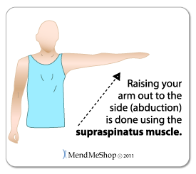 The supraspinatus is the most frequently torn of all the rotator cuff tendons. It is the uppermost muscle of the rotator cuff and is located at the back of your shoulder blade. It passes beneath the acromion and runs towards the greater tubercle at the top of your humerus joining at the top of the cuff by the supraspinatus tendon. Your supraspinatus muscle primarily helps you to bring your arm directly out to the side(known as abduction) although it assists you with other shoulder motions as well. 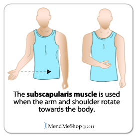 The subscapularis is the largest and the strongest of all your rotator cuff muscles. It completely covers the front of the shoulder blade. This muscle is attached to the front of the humerus which allows you to move your upper arm inward toward the center of your body (known as internal rotation). The infraspinatus muscle is located at the back running from the bottom of the shoulder blade across to the top of the humerus. This muscle works with the teres minor muscle to move your arm outward, away from the center of your body (known as external rotation). 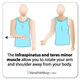 The teres minor muscle sits below the infraspinatus and runs at the same angle attaching just below the greater tubercle of your humerus bone. The teres minor also assists with the outward rotation of your arm from the center of your body (know as external rotation). Other main muscles in the shoulder area include:
Shoulder Bones and LigamentsThe bone structures inside the shoulder that are significant to the rotator cuff include the humerus, the scapula, the acromion process, the clavicle, the greater tubercle, and the glenoid cavity. 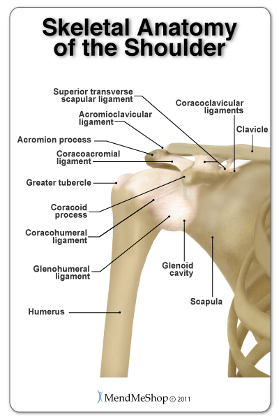 The humerus (upper arm bone) runs from your elbow to your shoulder and meets at the rotator cuff with a ball-like end known as the greater tubercle. This is the 'ball' part of the ball and socket joint in your shoulder.
The scapula (shoulder blade) is a triangular shaped bone with 2 bony projections at the top, right at your shoulder cuff. One of these projections is referred to as the acromion and it sits above the humerus. The other is called the coracoid process and it sits in front of the acromion and below the clavicle. Where your humerus meets your scapula there is a very shallow concave 'socket' known as the glenoid cavity (also called the glenoid fossa). Ligaments are soft tissue bands that connect one bone to another. The joints of the shoulder that are primarily responsible for movement are held together by several strong ligaments. They include the coracoclavicular ligaments, the coracoacromial ligaments, the superior transverse scapular ligament, the coracohumeral ligament, the acromioclavicular ligament, and the glenohumeral ligaments. Shoulder JointsInside the shoulder there are three joints; the glenohumeral joint, the acromioclavicular joint (A/C joint) and the sternoclavicular joint. The glenohumeral joint is a joint where the greater tubercle (humeral head at the top of the arm bone) meets the shoulder socket of the scapula, called the glenoid cavity or glenoid fossa. Inside the joint, the labrum (a form of cartilage) cushions the humeral head against the glenoid. 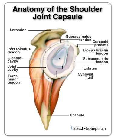 This joint is considered a ball and socket joint however the 'socket' is not as deep as similar joints in your body. Instead, the humerus sits against the glenoid cavity similar to how a golf ball sits on a tee. Since the ball does not fit directly inside socket of the glenohumeral joint, it is the labrum, muscles, and tendons that hold the ball of the humerus against the glenoid fossa providing stability between your scapula and your humerus. Due to this shallow socket and the scapula 'floating' above the rib cage (connected to the clavicle by ligaments, muscles and tendons) your shoulder is able to move around freely in several directions. This makes the shoulder the most mobile joint in the body. However, this also makes it the least stable joint and the one most prone to injury. The acromioclavicular joint (A/C joint) is a gliding joint between the clavicle and the acromion. The acromion is a bony projection that comes off the scapula and forms the point at the outside edge of your shoulder. The acromioclavicular joint allows you to rise your arm over your head. If this joint dislocates it is commonly known as a separated shoulder. The sternoclavicular joint, where the collar bone meets the sternum (breast bone) is not considered as important for shoulder movement. Bursae in the ShoulderIn your shoulder joint there are 4 main bursae; the subacromial bursa, the subcoracoid bursa, the subscapular bursa, and the subdeltoid bursa. 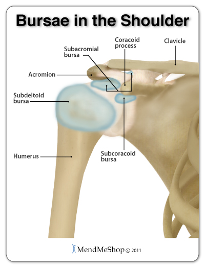 Bursae (plural for bursa) are fluid filled sacs that act as cushions to help the bones and soft tissue move smoothly within the joint. Your bursae also act as padding to protect your soft tissue from the bony points on the scapula, coracoid, and acromion. The subacromial bursa is the most susceptible to bursitis in the shoulder. It's located in the subacromial space, between the acromion and the humeral head (greater tubercle), and is used frequently during shoulder movement to reduce friction. The risk of impingement of this bursa in the subacromial space is high because the area is small. What Causes a Rotator Cuff Injury?Rotator cuff injuries are very common, especially in people over 40 years of age. Most problems involve damage and irritation to the rotator cuff soft tissues (muscles, ligaments, tendons and bursa) rather than the bones, as they move frequently within a tight space. A rotator cuff injury usually begins as inflammation caused by some form of small but continuous source of irritation, such as repetitive overhead motions from sporting activities, work tasks or daily chores, which can lead to tendonitis, tendinosis, frozen shoulder, impingement, or bursitis. If you do not address the cause of the inflammation, a partial or complete tear (rupture) can develop in your rotator cuff due to chronic wear and tear of the tendon. A tear may also result at any age from an acute or single traumatic event, such as a fall onto an outstretched arm. Learn More About The Rotator CuffLearn more about Shoulder Surgery and Post-Surgery Recovery Learn more about The TShellz Wrap Learn more about which is better for your rotator injury - ice or heat FREE SHIPPING ON ALL PRODUCTS CURRENTLY ENABLED |
|

